 首页
首页 400-620-6333
Bielschowsky Silver Staining Technique
Licia Miller Product Manager
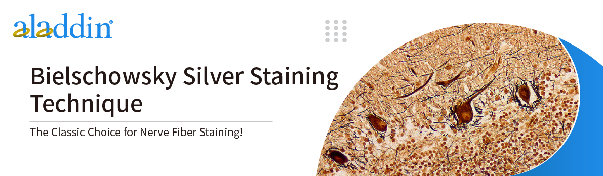
Bielschowsky silver staining is a classic silver staining technique, which is mainly used in the pathological study of nervous system tissues, especially in showing structures such as neuronal axons, neurofibrillary tangles and senile plaques.
This technique was developed by Max Bielschowsky, a famous neuropathologist in the early 20th century, based on the silver staining methods of predecessors (such as Ramón y Cajal and Fajersztajn), to develop a modified silver staining method suitable for formalin-fixed tissues. He treated tissue sections with silver nitrate and ammoniacal silver solutions and used reducing agents to selectively deposit silver particles in the axoplasm of nerve fibers, significantly improving the clarity and stability of staining.
This method achieved for the first time the effective staining of nerve fibers in old tissues (such as long-term fixed pathological specimens), solving the problem of early methods' dependence on fresh tissues.
In general, the staining principle of the Bielschowsky silver staining method is based on the reduction reaction of silver ions and the auxiliary effect of formaldehyde. During the staining process, formaldehyde first reacts with components such as proteins in nerve fibers, changing the chemical properties of the surface of nerve fibers, thereby providing conditions for the deposition of silver ions. Subsequently, silver ions are reduced to metallic silver particles under appropriate conditions, and these metallic silver particles will be deposited on the nerve fibers, making them appear black. This black metallic silver particle deposition effect allows the nerve fibers to be clearly observed under a microscope, thereby achieving specific staining of the nerve fibers.
Experimental Steps
1. Prepare the sections in advance, usually paraffin sections or frozen sections. The thickness of the sections is generally 10-20 microns, which can be adjusted according to experimental requirements and tissue type.
2. Hydrate the sections with dH2O (paraffin sections need to be dewaxed with xylene in advance). Then hydrate with different concentrations of ethanol in turn, and finally rinse with distilled water 3 times, 3 minutes each time.
3. Place the sections in 50 mL of 10% silver nitrate and incubate at 37°C in the dark for 25-35 minutes (the time may vary slightly for different kits).
NOTE: Temporarily keep this solution after incubation (for step 4).
4. Wash in distilled water 3 times, 3 minutes each time.
5. Add concentrated ammonium hydroxide dropwise to the silver nitrate solution retained in step 3 while stirring.
6. Place the sections in this solution and incubate at 37°C for 15 minutes. Reserve this solution for step 8.
7. Wash the sections three times with 0.1% ammonium hydroxide at room temperature for 2 minutes each time.
8. Add 350 μL of developer solution (0.2 mL 37% formaldehyde, 12 mL dH2O, 12.5 μL 20% nitric acid, and 0.05 g citric acid) to the silver hydroxide solution left over from step 6.
9. Place the sections in the solution and stain for 10 minutes until they turn black.
10. Wash the sections three times with 0.1% ammonium hydroxide for 2 minutes each time, and then wash them three times with distilled water for 2 minutes each time.
11. Incubate in 0.2% gold chloride for 5 minutes for color adjustment.
12. Add 5% sodium thiosulfate and fix for 1 minute.
13. Wash with distilled water, dehydrate in alcohol, then dehydrate in xylene and mount the slides.
Application Fields
1. Diagnosis of nervous system tumors
Used to differentiate neurofibroma (positive) from schwannoma (negative) and to assist in pathological classification.
2. Neurodegenerative disease research
Pathological features such as neurofibrillary tangles and senile plaques in the brain tissue of Alzheimer's patients can be clearly displayed through this staining.
3. Basic neuroscience research
It displays the neurofilament network and axon-dendrite structure in the neuronal cytoplasm, which is suitable for the study of neural development and injury repair.
Precautions
1. Experimental Condition Control
Avoid light: Silver solution is light-sensitive and needs to be kept away from light throughout the process to avoid nonspecific precipitation.
Temperature and time: The temperature (usually 37°C) and time of the silver immersion and development steps must be strictly controlled to prevent over-staining.
2. Reagents and Utensils
Use high-purity distilled water and clean glassware thoroughly to prevent contamination.
Some reagents (such as silver nitrate and ammonia) are corrosive and toxic and must be operated in a fume hood and wearing protective equipment.
3. Interpretation of results
Positive result: The nerve fibers appear dark purple to black, with a light yellow or brown background.
If there is poor uniformity or the background is too dark, it may be caused by insufficient reduction or incomplete cleaning.
For more product details, please visit Aladdin Scientific website.